CT – Computed Tomography
November 23, 2022
Equine veterinary hospital Comments Off on Standing tieback surgery for ‘roarers’
22
15
18
What is CT?
- The CT moves 360 degrees around the region of interest, taking multiple x-rays at various angles
- The images are then reconstructed to create a 3D image of an individual structure.
When is CT used?
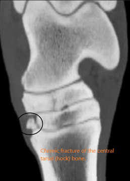
- Gold standard for bone changes (especially subtle bone changes)
- Bone chips and fracture lines
- Penetrating injuries, especially to the foot
- Surgical planning of fracture repairs
- Areas of complex anatomy (skull, sinuses, dentition, hyoid).
What are the benefits?
- No superimposition as with 2d x-rays – can reformat images in alternate cross-sectional planes/3D
- Very quick acquisition time, however preparation and positioning of the patient takes time.
What is the procedure?
- Full lameness evaluation to localise the region of lameness
- For standing CT, a catheter is generally placed to maintain sedation throughout
- Some CTs require a general anaesthetic
What are the limitations?
- Restricted to limbs, head and neck depending on the size of the patient and the CT bore
- MRI is still gold standard for soft tissue injuries. However, contrast agents can be used to highlight some soft tissue injuries
- Cost
- Availability
What are the risks?
- Minimal risk for standing CT
- Recovery from GA for a horse is always high risk
- Radiation safety applies in the same way as taking x-rays – handlers must wear a lead gown, thyroid collar and dosimeter. Ideally, a lead shield is placed between the handler and the patient.
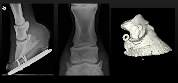
Reconstructed 3D image of the hoof

0 Comments