Equine Gastric Ulcer Syndrome (EGUS)
June 8, 2021
Equine veterinary hospital Comments Off on Standing tieback surgery for ‘roarers’
22
15
18
Introduction
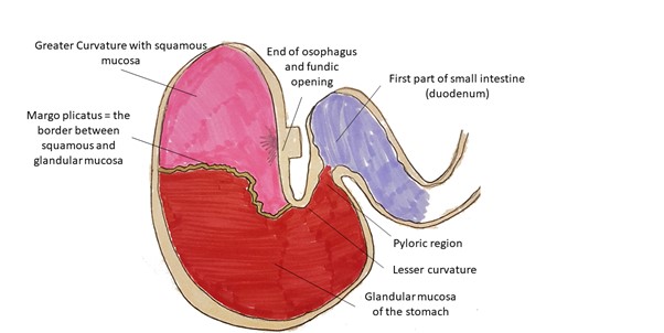
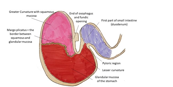
Equine Gastric Ulcer Syndrome (EGUS) is a very common gastrointestinal disease affecting many horses. Horses’ stomach is divided into two slightly different sections based on the mucosal type within the stomach. One section is the “squamous” section, and another is the “glandular” section.
Diagnosis of EGUS
How can we diagnose EGUS in an equine patient?
- Signalment and History:
Classically, horses with EGUS will present with typical clinical signs of low-grade abdominal discomfort (especially after food), we hear about the complaints of teeth grinding, inappetence, flank watching, being irritated when saddled up and also mild weight loss and loss of healthy hair coat. However, throughout my years of consulting for horses that are eventually diagnosed with EGUS, I have observed that the clinical signs for some horses may be rather insidious, including, reluctance to work, poor performance in racehorses, beating up paddock mates, being grumpy and irritable/sour, or unexplained weight loss. Some of these horses never displayed signs of inappetence (many are in very good body condition!) colic, or weight loss, or abdominal pain. Hence the suspicion of EGUS should be there if an equine patient presents with these clinical complaints.
- Diagnostic tests:
The definitive way to diagnose EGUS is by video gastroscopy. With a video gastroscopy, we can actually visualise the equine stomach from the cardia to the pylorus to rule in/out the presence of equine gastric ulcer syndrome.
Gastroscopy is a procedure where we pass a video endoscope into the stomach for visualisation. The procedure is not dissimilar to gastroscopy in humans where preparation in the form of fasting is required. In the horse, we perform this standing sedated while in humans the procedure is performed under GA.
- Preparation for gastroscope;
To prepare your horse for a gastroscopy, he/she will have to be fasted just like in humans. My protocol for preparation is to provide dinner to the horse, as usual, the day before but remove all feed material by 10 pm. This also means that the horse should be in a stable with only water and not in a paddock as they will still graze grass. Water would then be removed from the stable at midnight. The procedure will then be performed the next day around 9 am giving a good fast to ensure that the stomach is as empty as possible. An empty stomach will allow us to visualise the mucosa and structures of the stomach without obstructions. We can either do this preparation in the hospital with an overnight stay or if the clients have a stable at home this preparation can also be done at home.
The procedure of gastroscopy is done standing sedated. We administer a sedative through the vein and the video scope is passed into the stomach via the nostril, just as one would pass a stomach tube for drenching, except this time, we can visualise the entire procedure.
The scope will enter the stomach via the oesophagus, and we first encounter the area called the greater curvature of the stomach. Here we can visualise most of the squamous mucosa and the border between squamous and glandular mucosa. This border is called the margo plicatus and is an area prone to ulceration. Following that, the scope is manoeuvred in a pathway that allows the scope to visualise the cardia and the lesser curvature of the stomach. The scope is then advanced into the pyloric region and this is the entrance to the first section of the small intestines. The pylorus is also an area that is prone to glandular ulcers.
Here are some clinical pictures of equine gastric and glandular ulcers, diagnosed at the Camden Equine Centre.
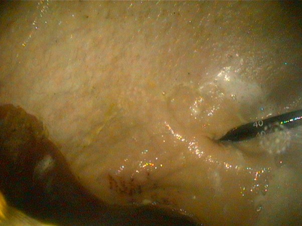
Image 1. Superficial squamous ulcers near the cardia of the stomach (cardia is the first closeding into the stomach after the oesophagus – see the scope emerging from the cardia on the right of the picture). Grade 2/5 EGUS.
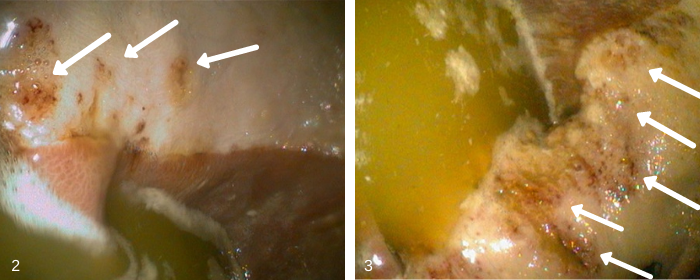
Images 2 & 3 – An extensive “row” of moderately deep squamous ulcers along the margo plicatus of a horse with weight loss and cranky behaviour. Grade 3/5 EGUS
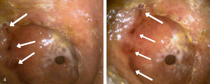
Images 4 & 5 – These images show several areas of moderately severe glandular ulcers at the pyloric closeding. Pyloric closeding is the “hole” in the middle of the picture. Pyloric closeding leads to the first section of the small intestines. This horse showed no weight loss or appetite loss but had two episodes of buckling at the knee assumed to be sudden pain.
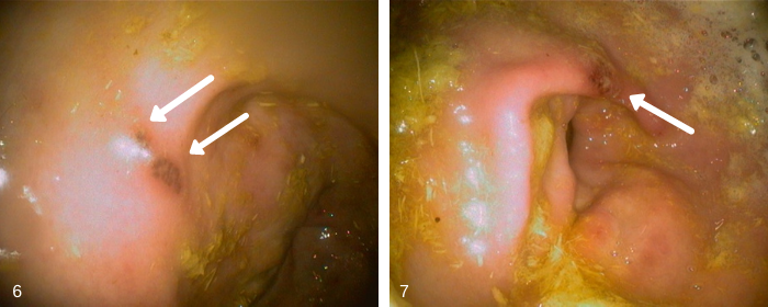
Images 6 & 7 – These 2 pictures are of a single focal area of moderately severe glandular ulcer near the closeding of the pylorus. This horse showed signs of aggression when being saddled and was cranky at work. No weight loss or inappetence noticed.
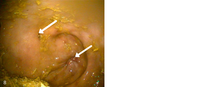
Image 8 – This picture shows several areas of moderately severe glandular ulcers at the pyloric closeding. This horse showed no weight loss however was girthy and cranky when saddled.
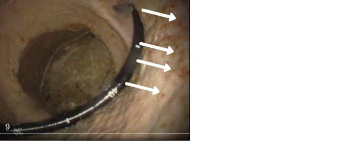
Image 9 – This horse had multiple pinpoint superficial ulcers peppered throughout the greater curvature of the stomach. Classified as Grade 1-2/5 EGUS.
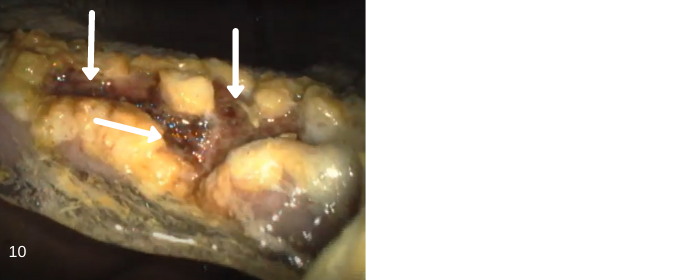
Image 10. The same horse pictured above, with an extremely severe ulcerated area of the lesser curvature. Classified as Grade 4/5 EGUS in this area based on depth and presence of clotted blood. This also appears to be chronic as there are areas of hyperkeratosis (scabbing) noted around the ulcer (seen as the yellow thickened area surrounding the ulcerated portions). Visualisation of the lesser curvature and pylorus cannot be achieved without retroflexing the scope. This horse showed signs of mild intermittent colic without any weight loss.
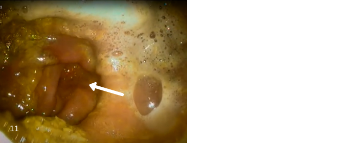
Image 11 – Generalised hyperaemic (increased in redness) of the entire pylorus area in this horse with clinical signs of being very cranky.
Treatment of EGUS:
The pharmacological treatment of EGUS is with the use of medications that reduce the production of acid in the stomach. The most used medications are omeprazole and ranitidine. Omeprazole remains the most popular as it is very sustainable at a once a day oral/in-feed administration. Fact: Humans also use omeprazole or esomeprazole as a treatment for gastric ulcers/heartburn and you can buy those over the counter at the pharmacy. The minimum course to treat a diagnosed gastric ulcer via endoscopy is at least 28 days, where a treatment dose is initiated for 2 weeks and a maintenance dose for a further 2 weeks.
Occasionally, some horses will require an extension past the 4-week therapy. A repeat gastroscopy to check for positive progression or resolution is highly recommended to determine if an extension of the therapy is required. Some horses will also require periodic treatment of gastric ulcers based on the specific “triggers” that cause ulcers in individual patients.
Dietary management by providing constant access to pasture or hay to digest will reduce the risk of gastric ulceration. Constant access to pasture or hay encourages the production of saliva and this in turn buffers the acidity of the stomach. Breaking up concentrates to smaller feeds throughout the day is also a good way to reduce the risk of gastric ulcers.
Written by DR ELIZABETH S.Y TEE DVM GradDip (VetClinStud) GradCert (HigherEd) FHEA FACVSc | Registered Specialist in Equine Internal Medicine

0 Comments